- صفحه نخست |
- سفارش کتاب |
- چاپ کتاب |
- فروشگاه |
- اخبار |
- درباره ما |
- ارتباط با ما |
- عضویت در سایت |
- ورود به سایت |
- ویدئوها
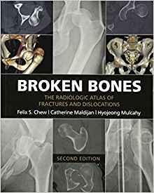
جزییات کتاب Broken Bones: The Radiologic Atlas of Fractures..2016
طبقه : اورتوپدی
استخوان های شکسته: اطلس رادیولوژیک شکستگی ها
Broken Bones: The Radiologic Atlas of Fractures..2016
مشخصات کتاب
ISBN (شابک): | ISBN-13: 978-1107499232 ISBN-10: 1107499232 |
قطع: | رحلی |
ناشر: | |
تعداد صفحات: | 406 |
سال و نوبت چاپ: | 2nd Edition 2016 |
نوع جلد: | هارد |
قیمت خرید کتاب : 899,200 تومان
جزییات بیشتر :
Broken Bones contains 434 individual cases and 1,101 radiologic images illustrating the typical and less typical appearances of fractures and dislocations throughout the body. The first chapter describes fractures and dislocations of the fingers, starting with fractures of the phalangeal tufts and progressing through the distal, middle, and proximal phalanges and the DIP and PIP joints. Subsequent chapters cover the metacarpals, the carpal bones, the radius and ulna, the elbow and upper arm, and the shoulder and thoracic cage. The cervical spine and the thoracic and lumbosacral spine are covered in separate chapters, followed by the pelvis, the femur, the knee and lower leg, the ankle, the tarsal bones, and the metatarsals and toes. The final three chapters cover the face, fractures and dislocations in children, and fractures and dislocations caused by bullets and nonmilitary blasts.
بازدید : 2335 مرتبه
محصولات مشابه
ناشرین
Elsevier (640) LWW (141) McGraw-Hill Education (106) Springer (97) تیمورزاده نوین (79) Thieme (67) Wiley-Blackwell (60) CRC Press (55) Academic Press (43) Oxford University Press (37) Cambridge University Press (32) Kaplan Publishing (24) Saunders (24) McGraw Hill / Medical (21) American Academy of Ophthalmology (17) Mosby (16) Elsevier (16) Jaypee (15) Jones & Bartlett Learning (15) Pearson (12) American Academy (12) lww (11)

